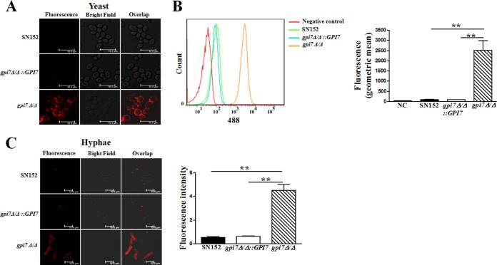
FIG 2.
Cell wall β-(1,3)-glucan was highly exposed in the C. albicans gpi7 mutant in both yeast and hyphal forms. Yeast-form growing cells (A and B) and hyphal growing cells (C) were stained with anti-β-(1,3)-glucan primary antibody to visualize β-(1,3)-glucan. The fluorescence intensity was quantified by flow cytometry (B, right) and the Leica LAS AF Lite program (C, right). The scale bar represents 10 μm. Data represent means (± SD) from triplicates of one representative experiment of three. **, P < 0.01 (one-way ANOVA with Bonferroni's posttest).

