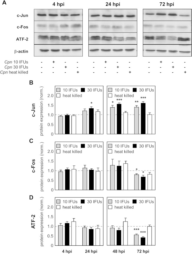
FIG 1.
Differential regulation of AP-1 proteins after C. pneumoniae infection. Lysates from uninfected HEp-2 cells (control), HEp-2 cells infected with 10 IFUs (low dose) or 30 IFUs (high dose) of C. pneumoniae (Cpn), or HEp-2 cells infected with heat-killed C. pneumoniae were prepared at various time points after infection (4 hpi, 24 hpi, 48 hpi, and 72 hpi). (A) Western blot of the indicated proteins at 4 hpi, 24 hpi, and 72 hpi. (B to D) Using densitometry, expression of the c-Jun (B), c-Fos (C), and ATF-2 (D) proteins was quantified during C. pneumoniae infection of HEp-2 cells. Data were normalized (norm.) against those for the uninfected control (dashed gray lines). Data, presented as the mean ± SD, and immunoblots are representative of those from at least four independent experiments (n = 4 to 6). *, P < 0.05; **, P < 0.01; ***, P < 0.001.

