FIG 5.

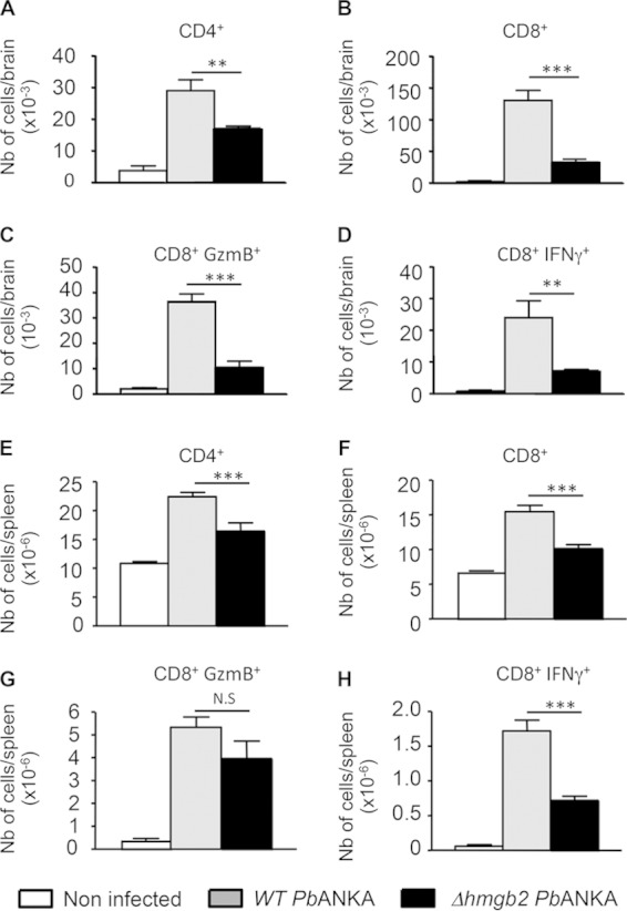
Reduced infiltration and reduced activation of CD4+ and CD8+ T cells in the brains of Δhmgb2 ANKA-infected mice. At the coma stage (day 6 postinfection), brains from WT- or Δhmgb2 ANKA-infected C57BL/6 mice, with 105 infected erythrocytes per mouse, were taken, and leukocytes associated with cerebral tissue were analyzed by FACS for the presence of the indicated leukocytes, expressed as absolute numbers per brain. Six mice per group were used. Values represent the means ± SD for one experiment of three. *, P < 0.05; **, P < 0.01; ***, P < 0.001; N.S, not significant. (A to D) CD4+, CD8+, CD8+ GzmB+, and CD8+ IFN-γ+ cells in brains. (E to H) The same cells were also analyzed in spleens. Multiple comparisons of brain-infiltrating T cells were analyzed by the Kruskal-Wallis test (with Dunn's posttest).
