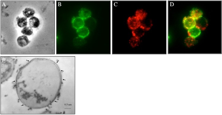
FIG 1.

β-1,3 and β-1,6 glucans are present in the outer cell wall layer of P. carinii. (A) Phase-contrast microscopy image of P. carinii cyst forms. (B and C) P. carinii cysts were fixed, and antibodies specific for β-1,3 glucan (B) and β-1,6 glucan (C) were used to localize the respective carbohydrates. (D) Merged β-1,3 glucan and β-1,6 glucan localization. (E) Representative transmission immune electron micrograph (n = 20) utilizing 18-nm gold particles showing labeling of β-1,6 glucans in the P. carinii cyst wall (arrows).
