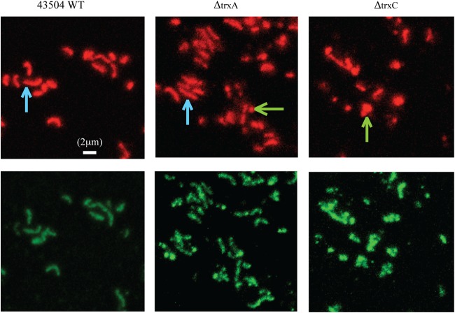
FIG 4.
Detection of 8-oxoG by immunofluorescent staining. Wild-type H. pylori, ΔtrxA mutant, and ΔtrxC mutant cells were fixed on a glass slide and stained with 8-oxoG-specific avidin-FITC conjugate (lower panel) and propidium iodide (upper panel), followed by examination via fluorescence microscopy. The contrast adjustment was normalized for all the images, and a representative set of images is shown here. Blue arrows point to some examples of bacillary cells in the wild-type and ΔtrxA strains, and green arrows highlight some coccoid or broken cells in the ΔtrxA and ΔtrxC mutant strains. A 2-μm bar is given as a size scale.

