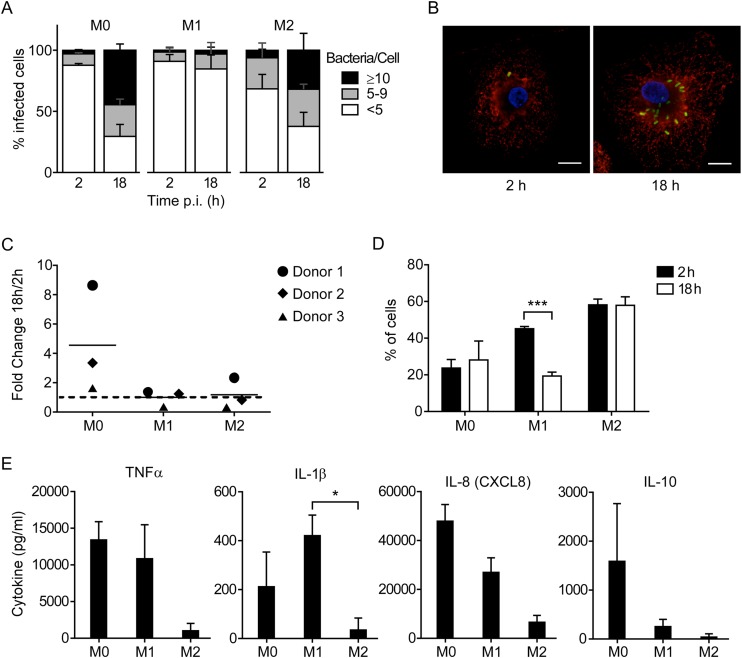
FIG 1.
Cell phenotype affects the ability of Salmonella Typhimurium to survive and replicate in human MDM. (A) M1, M2, and M0 macrophages were infected with wild-type strain SL1344, fixed, and stained for immunofluorescence microscopy at the indicated times. Infected cells were scored as containing ≥10, 5 to 9, or <5 bacteria. The means ± standard deviations (SD) from 3 independent experiments using three different donors are shown. (B) Representative images of infected M0 macrophages. Green, LPS; red, LAMP1; blue, DAPI. Bar, 10 μm. (C) Gentamicin protection assays were performed using cells infected as described for panel A. At 2 and 18 h p.i., the cells were lysed and recoverable CFU were assessed by plating. Each point represents the fold change in CFU (18 versus 2 h p.i.) obtained in one experiment, and lines represent the means from 3 independent experiments. (D) Cells were infected, fixed, and stained for microscopy as described for panel A. The percentage of cells containing bacteria at each time point is shown. Bars represent means ± SD from 3 independent experiments. ***, P = 0.0003 by two-way ANOVA. (E) Cytokine levels in cell culture supernatants at 12 h p.i. Results are means ± SD from 3 independent experiments. *, P = 0.027 by one-way ANOVA.

