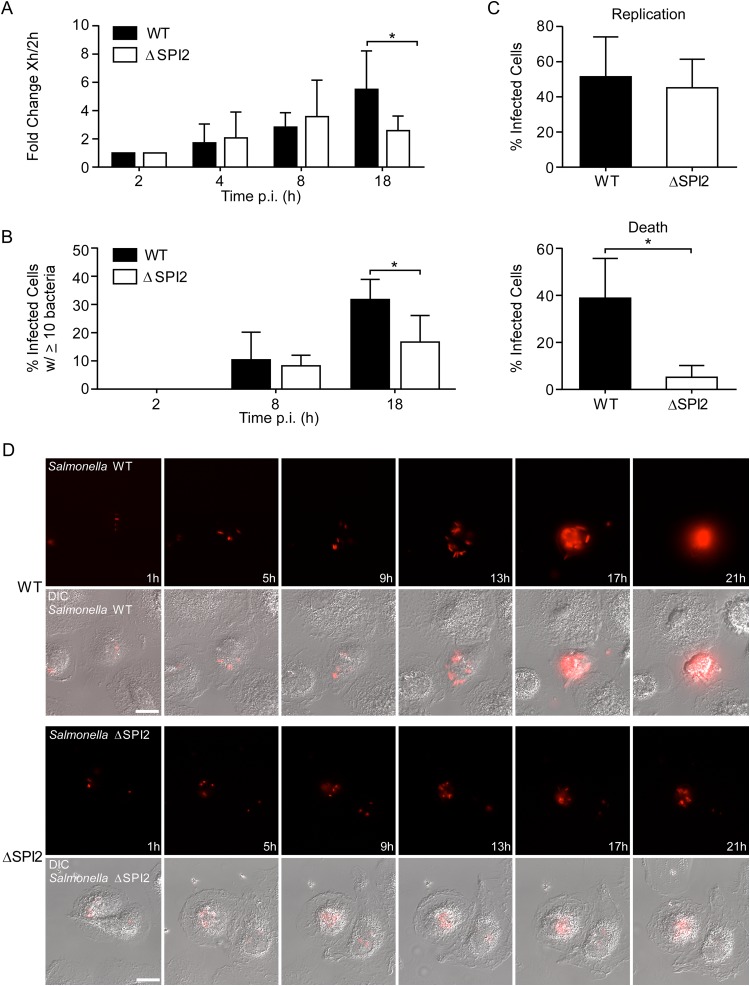
FIG 6.
SPI2-dependent and -independent replication of Salmonella Typhimurium in M0 macrophages. (A) Gentamicin assay. Shown is the fold change in recoverable CFU compared to the values at 2 h p.i. Results are means ± SD from 5 independent experiments. (B) The percentage of infected cells containing the indicated number of bacteria was assessed by microscopy. Results are means ± SD from 3 independent experiments. (C and D) Intracellular replication was assessed by live cell imaging. Macrophages infected with mCherry-expressing WT or ΔSPI2 bacteria were imaged from 1 to 20 h p.i. every 15 min. (C) Cells that contained bacteria at 1 h were assessed for bacterial replication (top) and the appearance of cell death by morphological changes (bottom) over the time course. Macrophages from 3 donors were analyzed for each strain. A total of 62 cells infected with the WT and 59 infected with the ΔSPI2 strain were analyzed. (D) Stills from representative movies showing the replication of mCherry-expressing WT (top) or ΔSPI2 (bottom) bacteria in macrophages. Also see Movies S1 and S2 in the supplemental material. Bar, 10 μm. *, P < 0.05 by two-way ANOVA (A and B) and by paired t test (C).

