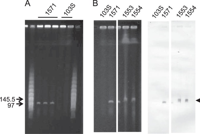
FIG 1.

Detection of pVAPN by PFGE. (A) Genomic DNA of bovine isolate 1571 and equine isolate 103S; three and two independent lysates per strain are shown. Relevant positions of the lambda PFGE marker (New England BioLabs) are indicated. pVAPN is observable as a distinct band of ≈100 kb in the bovine isolate. (B) Southern blot analysis of bovine isolates PAM1571, PAM1533, and PAM1554 (strain 103S was used as a negative control). (Left) Relevant sections of the PFGE gel; (right) membrane hybridized with a pVAPN-specific DNA probe (600-bp fragment encompassing the 3′ region of vapN and the 5′ region of vapQ). The arrow indicates the pVAPN band.
