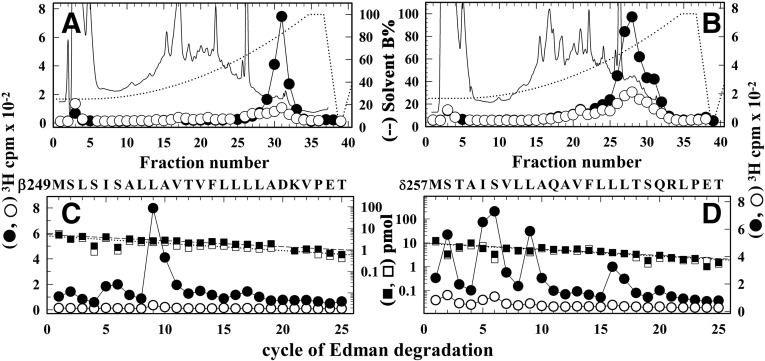
Fig. 6.
Agonist-enhanced [3H]dFBr photolabeling in βM2, and δM2. Torpedo nAChR-rich membranes were photolabeled at preparative scale with 0.5 µM [3H]dFBr in the absence (○, ⬜) or presence (⬤, ▪) of 1 mM Carb. (A and B) Elution of peptides (solid line) and 3H (○, ⬤) during rpHPLC purifications of an ∼8 kDa β subunit fragment isolated by from a trypsin digest (A) and an ∼14 kDa δ subunit fragment isolated from an EndoLys-C digest (B). (C and D) 3H (○, ⬤) and phenylthiohydantoin amino acids (⬜, ▪) released during sequence analyses of rpHPLC fractions 30 to 31 from (A) and fractions 27–29 from (B), respectively. (C) The only sequence detected began at the N-terminus of βM2 (βMet249; 5 pmol each condition), and the major peak of 3H release in cycle 9 indicates photolabeling of βLeu257 (βM2-9; −Carb/+Carb, 2/50 cpm/pmol). (D) The only sequence detected began at the N-terminus of δM2 (δMet257; 10 pmol each condition). The peaks of 3H release in cycles 2, 5, 6, 9, and 16 indicate photolabeling (−Carb/+Carb, in cpm/pmol) at δSer258 (δM2-2; 1/7), δIle261 (δM2-5; 1/14), δSer262 (δM2-6; 2/17), δLeu265 (δM2-9; 0.5/14), and δLeu272 (δM2-16; 1/12).

