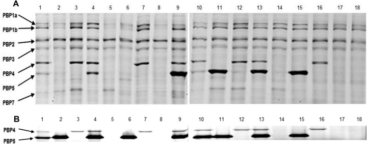
FIG 1.

Bocillin FL binding test of PAO1 wild-type and derived mutants. (A) Conventional cell membrane preparation protocol. (B) Modified protocol to avoid AmpC contamination of cell membrane preparations leading to Bocillin FL hydrolysis. The PBP pattern (at left) of all the constructed P. aeruginosa mutants and the wild-type PAO1 (lanes 1 to 18) were visualized by fluorescence scanning using the Typhoon 9410 variable-mode imager at 588 nm, with a 520 BP 40 emission filter, after an SDS-PAGE run of the reaction samples in 8% acrylamide gels, in which each reaction involved an incubation of 100 μg of cell membrane protein with 10 μM Bocillin FL at 37°C for 30 min. Lanes 1 and 10, wild-type PAO1; lane 2, PAO ΔdacB; lane 3, PAO ΔdacC; lane 4, PAO ΔpbpG; lane 5, PAO ΔdacB ΔdacC; lane 6, PAO ΔdacB ΔpbpG; lane 7, PAO ΔdacC ΔpbpG; lane 8, PAO ΔdacB ΔdacC ΔpbpG; lane 9, PAO ΔampC; lane 11, PAO ΔdacB ΔampC; lane 12, PAO ΔdacC ΔampC; lane 13, PAO ΔpbpG ΔampC; lane 14, PAO ΔdacB ΔdacC ΔampC; lane 15, PAO ΔdacB ΔpbpG ΔampC; lane 16, PAO ΔdacC ΔpbpG ΔampC; lane 17, PAO ΔdacB ΔpbpG ΔampC ΔdacC; and lane 18, PAO ΔdacB ΔdacC ΔpbpG ΔampC.
