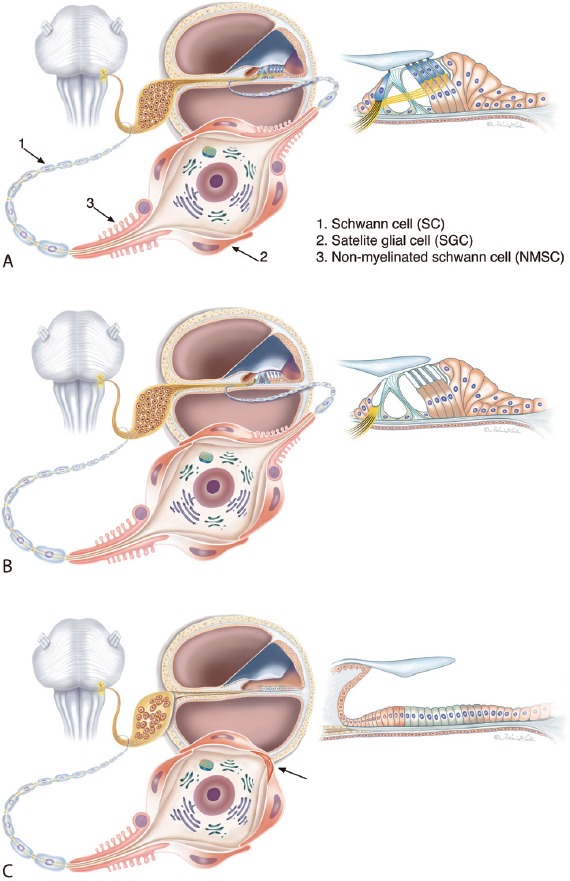
Figure 1.

Illustration of different cell anatomies in patients with sensorineural deafness.
(A) Normal condition. The spiral ganglion neurons (SGNs) are surrounded by satellite glial cells (SGCs) while the pre- and post-somatic axonal segments are bordered by non-myelinated Schwann cells (NMSC). Axons are enwrapped by regular Schwann cells. (B) Deafness associated with loss of hair cells. Preservation of supporting cells maintains the integrity of the peripheral dendrite. (C) Atrophy of the sensory epithelium results in dendrite degeneration. The bordering cells (SGCs and NMSCs) consolidate neurons as mono-polar or “amputated” cells (arrow) with unbroken connections to the brain stem. Theoretically, these neurons could be re-sprouted. This condition could be prevalent in patients with long deafness duration (Modified from Liu et al., Neuroscience. 2015;284:470-482). Graphic Karin Lodin.
