Figure 2.

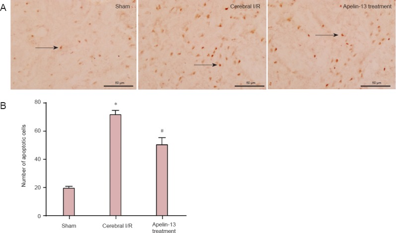
Apelin-13 reduced apoptosis in the ischemic penumbra region in a middle cerebral artery occlusion rat model after 2 hours of ischemia and 24 hours of reperfusion, as detected by TdT-mediated dUTP nick-end labeling (TUNEL) staining.
(A) Representative photomicrographs of apoptotic cells with TUNEL in sham, cerebral I/R, and Apelin-13 treatment groups (fluorescence microscope, × 400, scale bars: 50 μm). Arrows indicate TUNEL positive cells. (B) Optical density values were used to calculate apoptotic cell number. *P < 0.01, vs. sham group; #P < 0.05, vs. cerebral I/R group (one-way analysis of variance followed by the least significance difference test). Data are expressed as the mean ± SD (n = 3 for each group). I/R: Ischemia/reperfusion.
