Fig. 3.
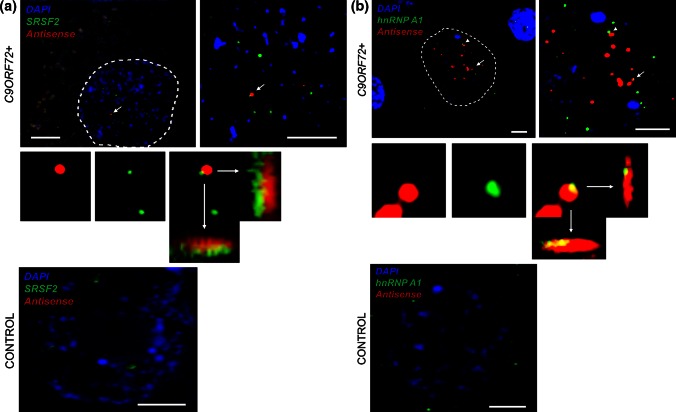
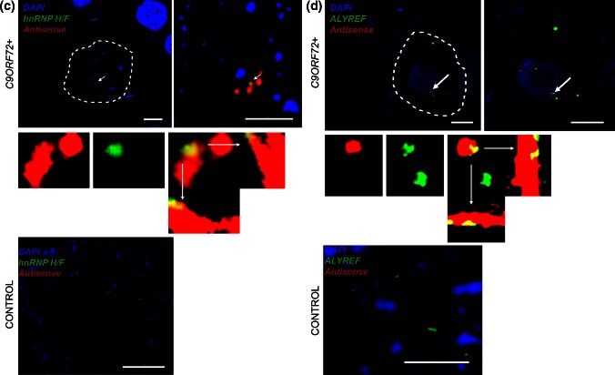
Combined RNA FISH and IHC demonstrate colocalisation of nucleolin and nuclear speckle components with antisense RNA foci in Purkinje neurons from C9ORF72-ALS patients and the distribution of these proteins in Purkinje neurons from control individuals. SRSF2 (a), hnRNP A1 (b), hnRNP H/F (c), ALYREF (d), and hnRNP K (e) are observed to colocalise with antisense RNA foci (arrows) in Purkinje neurons from C9ORF72-ALS patients. A large scale view is shown to the left of a zoomed-in image. Colocalisation events are enlarged including orthogonal views, and unmerged protein and RNA foci are shown for comparison. There was not a significant difference between the staining of these proteins in controls and C9ORF72+ individuals, but no antisense RNA foci are observed in controls. Nucleolin was not observed to colocalise with antisense RNA foci (f); moreover, the distribution of nucleolin was variable in C9ORF72+ Purkinje neurons. In some cells, nucleolin was prominently nucleolar (f, left panel) and in other cells it was dispersed throughout the nucleus (f, right panel, RNA focus is indicated by an arrowhead). The dotted line illustrates the nuclear border in images a–e and the nucleolar border in image f. Scale bar 3 µm


