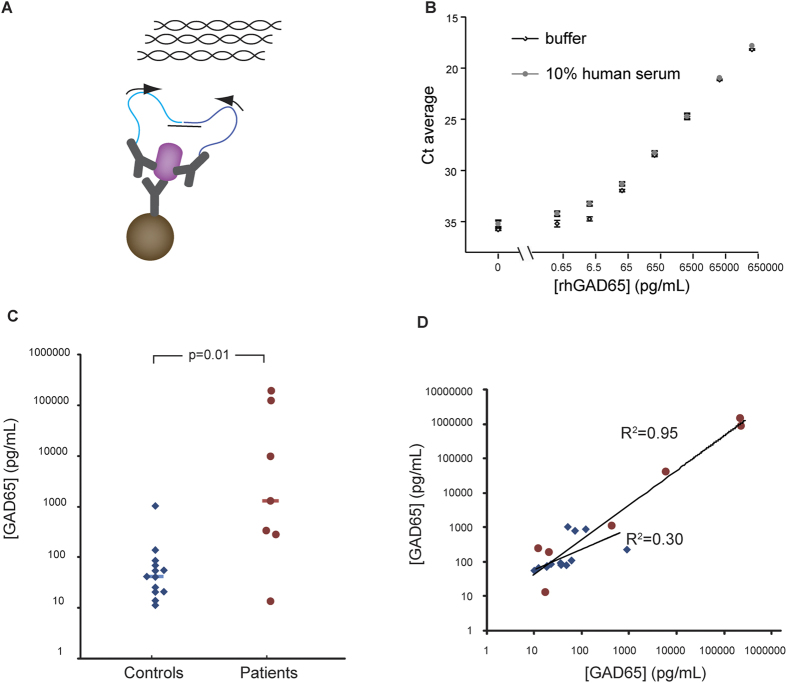
Figure 1. Detection of GAD65 by solid-phase PLA.
A) Goat polyclonal anti-human GAD65 antibodies were covalently coupled to magnetic beads for capturing GAD65 proteins from biological samples. Two other portions from the same anti-GAD65 polyclonal antibody preparation with distinct attached oligonucleotides, referred to as PLA probes, bound to GAD65 captured on the magnetic beads. Upon hybridization of a connector oligonucleotide the two antibody-conjugated DNA oligonucleotides were enzymatically joined to serve as an amplification template, and quantified by qPCR. B) Solid-phase PLA was used to detect recombinant human GAD65 (rhGAD65) in a dilution series (65 ng/ml – 0.65 pg/ml) in 10% control human serum and assay buffer. The limit of detection was approximately 0.65 pg/ml. Averages from triplicate measurements are shown with error bars that indicate the standard deviation. C) Levels of GAD65 were compared in serum samples from SPS patients and controls. Significantly increased GAD65 levels (p = 0.01) were observed for the SPS group (n = 7) compared to the controls (n = 13). The horizontal bars indicate the median for the patient and control groups. The assays were performed in three replicates. D) Scatter plot showing the correlation between two independent measurements of GAD65 in serum samples from SPS patients (R2 = 0.95) and controls (R2 = 0.30), red circles represent SPS patients and blue diamonds are controls.

