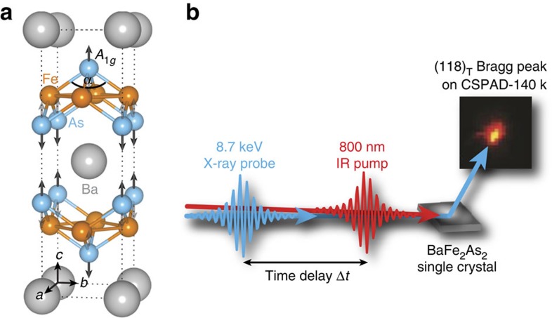
Figure 1. Crystal structure and time-resolved X-ray scattering.
(a) Tetragonal crystal structure of BaFe2As2 in the presence of the A1g phonon mode, parametrized by the Fe–As–Fe bond angle α. (b) Schematic of the experimental setup with the incoming infrared (IR) pump (red) and the X-ray probe pulse (blue). The temporal evolution of the diffraction pattern from the photo-excited BaFe2As2 single crystal was measured with a CSPAD-140k area detector. Δt is the time delay of the probe pulse with respect to the pump pulse.

