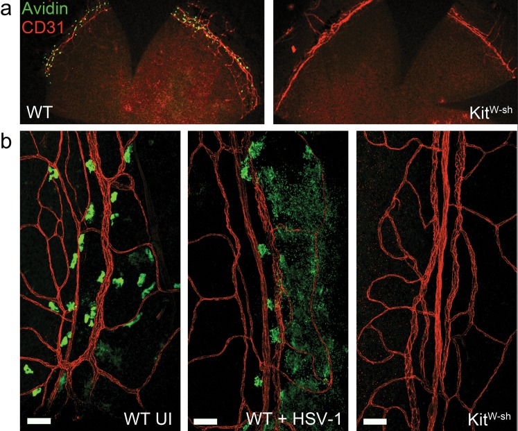
Figure 1.
Mast cells surround the cornea in the limbus and respond to corneal HSV-1 infection. (a) Corneolimbal button whole-mounts from WT and KitW-sh mice show MCs (green) near the CD31+ limbal vasculature (red). (b) Limbus-associated MC from uninfected (UI) WT mice have smooth, regular edges (left). Limbus-associated MC from WT mice at 24 hours pi with 1000 PFU HSV-1 per eye have a degranulated phenotype (center), and are absent in the tissue from KitW-sh mice (right). Scale bar: 50 μm. Images are representative of two independent experiments (n = 2–3 corneas per group). Representative images of limbus-associated MC were acquired using an Olympus MVX-10 epifluorescent microscope with ×16 zoom (a) or Olympus FV-500 confocal microscope with a ×20 objective (b).

