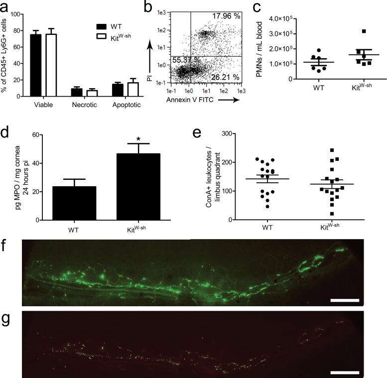
Figure 5.
Neutrophil characteristics and leukostasis in the limbus. Corneas were harvested from WT and KitW-sh mice at 24 hours pi, digested in type 1 collagenase, immunolabeled, and analyzed by flow cytometry for CD45+ Ly6G+ PMN with propidium iodide (PI) and annexin V staining for viability analysis (a). Results reflect % total PMN in five mice per group (two independent experiments). (b) Representative dot plot from a WT mouse showing PI and annexin V analysis: LLQ (PI− annexin V−) = viable; LRQ (PI− annexin V+) = apoptotic; URQ (PI+ annexin V+) = necrotic. (c) Neutrophils counts in peripheral blood at 48 hours pi (n = 6 mice per group; three independent experiments) evaluated by flow cytometric analysis as shown in Figure 3. (d) Total MPO was evaluated by ELISA at 24 hours pi (n = 9 samples/group; three independent experiments). Myeloperoxidase production was not detected in uninfected corneas of WT and KitW-sh mice (not shown). Total leukocytes adhering to the limbal vasculature in pericorneal quadrants 24 hours pi (e) visualized by ConA perfusion staining. Results show total leukocytes/quadrant representing 16 limbus quadrants from four corneas per group (three independent experiments). All data graphed as mean ± SEM. (f) Epifluorescent micrograph depicting ConA staining of vasculature (dim) and leukocytes (bright) in a limbal quadrant used for analysis in (e). (g) Color-intensity adjusted rendering of limbus in (f) to eliminate ConA+ vasculature for quantification of adherent ConA bright leukocytes. Images were obtained using an Olympus MVX-10 epifluorescent microscope at ×1.6 magnification (scale bar: 200 μm).

