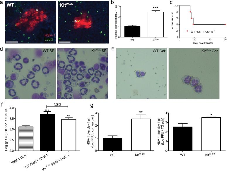
Figure 6.
Neutrophils facilitate HSV-1 replication through viral hijacking. (a) Confocal microscopy was used to visualize DAPI-stained nuclei (blue), HSV-1 antigen (red), and Ly6G+ PMN (green) in WT and KitW-sh cornea whole-mounts at 48 hours pi. Representative images of five corneas per group are shown (two to three independent experiments). Images were obtained using an Olympus FV-500 confocal microscope with a ×20 objective; images reflect additional ×3 digital zoom. Scale bar: 50 μm. Arrows point to regions of PMN and HSV-1 colocalization. (b) Real-time PCR analysis of HSV-1 TK relative to β-actin in Ly6G+ neutrophils immunomagnetically enriched from pooled WT or KitW-sh corneas harvested at 48 hours pi and digested in type I collagenase. Expression normalized to WT PMN. TK was detected in both WT- and KitW-sh-derived PMN. Data reflect two to four RT-PCR reactions on 2 × 10⁴ PMN isolated from pooled infected corneas of 6 to 10 mice per group (two independent experiments). (c) Survival of CD118−/− mice following intravenous adoptive transfer of 2 × 10⁴ PMN isolated from pooled WT corneas (12 mice) as in (b) delivered via retro-orbital injection (n = 6 CD118−/− mice; two independent experiments). Data reported as a percentage of survival of CD118−/− mice through day 30 post transfer. Each data point represents one CD118−/− mouse. Representative images shown in (d) and (e) are cytospin preparations of Diff-Quik stained Ly6G+ cells recovered from spleens (d) showing ring-shaped nuclear morphology and infected corneas (e) showing segmented nuclei at 48 hours pi (×96 magnification). (f) Ly6G+ cells (PMNs) isolated from peripheral blood were infected at 0.5 MOI with HSV-1 and evaluated for viral titer 18 hours pi to show that PMNs are sufficient for HSV-1 replication (n = 8–12 cultures containing 25,000 PMNs per culture; two independent experiments). A one-way ANOVA with Tukey's correction for multiple comparisons was used to calculate statistical differences in HSV-1 titers in infected PMN cultures over HSV-1 only control cultures. (g) Herpes simplex virus type-1 titer in the corneas and TG of WT and KitW-sh mice at day 4 pi (n = 5 animals/group; two independent experiments).

