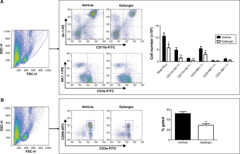
Figure 5.
Influence of galangin on recruitment and activation of inflammatory cells.
Notes: (A) IHLs were isolated from vehicle-pretreated CIH mice and galangin-pretreated CIH mice (2 hours after injection of Concanavalin A). The total number of IHLs from each mouse was counted using a cell counting plate. IHLs were stained with CD3e, CD11b, Gr-1, and NK1.1. The number of each cell subset was calculated by multiplying the total number of IHLs by the frequency of the subset. (B) IHLs were stained with CD3e and CD69, and the percentage of activated T-cells was analyzed. Data are shown as the mean ± standard deviation. The results are representative of three experiments. *P<0.05, **P<0.01.
Abbreviations: CIH, Concanavalin A-induced hepatitis; FITC, fluorescein isothiocyanate; APC, allophycocyanin; PE, R-phycoerythrin; SCC-H, side scatter-height; FSC-H, forward scatter-height; IHLs, intrahepatic leukocytes.

