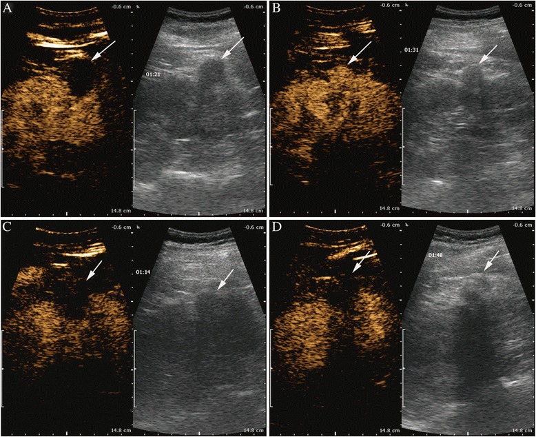
Fig. 2.

Sequential contrast-enhanced ultrasound images of the ablation area of renal cell carcinoma (RCC) observed for the period with two cryoablations. a Contrast-enhanced ultrasound 1 month after stage 1 cryotherapy. b Contrast-enhanced ultrasound 12 months after stage 1 cryotherapy. c Contrast-enhanced ultrasound 1 month after stage 2 cryotherapy. d Contrast-enhanced ultrasound 12 months after stage 2 cryotherapy
