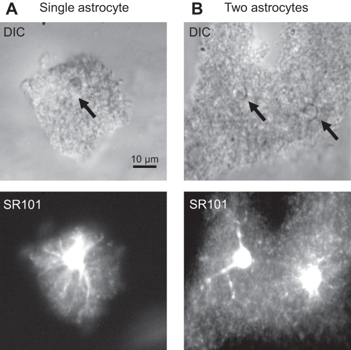
Fig. 1.

Morphology of astrocytes in freshly dissociated hippocampal tissues. A and B, top: differential interference contrast (DIC) images of 2 freshly dissociated hippocampal blocks. The block in A was identified as a single astrocyte based on the existence of a single, glial-like soma with diameter <10 μm and well-preserved domain territory, and the block in B contained 2 astrocytes whose identification was based on the same criteria (B). A and B, bottom: the initial morphological cell identification could be readily confirmed by the respective sulforhodamine-101 (SR-101) staining.
