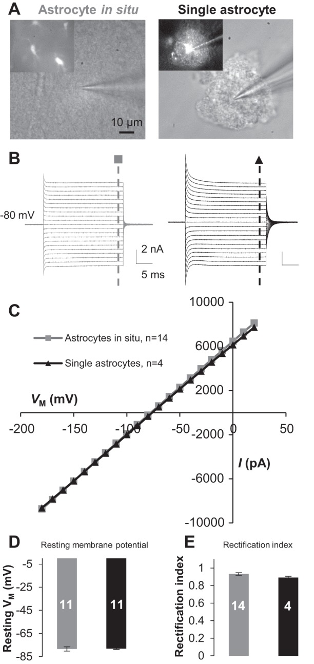
Fig. 2.

Freshly dissociated single astrocytes exhibit the same passive conductance and resting membrane potential (Vm) as astrocytes do in situ. A: DIC images were obtained during patch recording from a syncytial coupled astrocyte (left) and a single astrocyte in freshly dissociated tissue preparation (right). B and C: the astrocytes in A showed a similar passive membrane conductance (B) with comparable amplitudes in current-voltage (I-Vm) plots (C). D: the single astrocytes showed Vm comparable to that of the astrocytes in situ. E: the rectification index (RI) values were comparable between single and syncytial coupled astrocytes. Values are means ± SE; numerals on bars indicate the number of observations.
