Fig. 3.
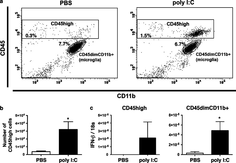
Poly I:C induced IFN-β in leukocytes. a A representative flow cytometry profile showing CD45high (leukocytes) and CD45dimCD11b+ (microglia) cell populations isolated from the CNS of mice treated with PBS or poly I:C, 6 h previously. b Number of CD45high cells isolated from mice treated with poly I:C (n = 5) compared to PBS (n = 3). c CD45high (leukocytes) and CD45dimCD11b+ cells (microglia) were sorted, pooled and IFN-β mRNA was measured by qRT-PCR. Bar graphs show IFN-β gene expression in sorted CD45dimCD11b+ microglia (n = 6) from poly I:C-treated mice compared to microglia (n = 6) from PBS-treated mice, in which IFN-β was also detected at a low level. Intrathecal poly I:C induced detectable IFN-β expression in sorted CD45high cells (n = 6), whereas IFN-β expression was not detected at all in sorted CD45high from PBS-treated mice (n = 6). Data were analyzed by two-tailed nonparametric Student’s t test followed by Mann–Whitney test. Results are presented as mean ± SEM. *P < 0.05

