
Fig. 4.
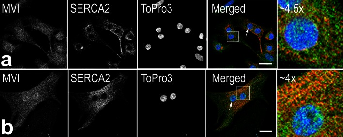
MVI colocalization with sarcoplasmic reticulum in neonatal rat cardiomyocytes. a and b, Localization of MVI (in green) and SERCA2 (in red) in day-2 and day-8 cardiomyocytes, respectively. Arrows point to the nuclear MVI presence. The far right panels, magnification (as marked in the figure) of the regions indicated in the merged panels. Nuclei were stained with TO-PRO®-3 (in blue). Bars 10 μm