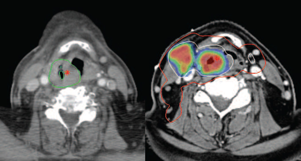
Figure 1.
Patient with a hypopharyngeal cancer (T3 N2c M0) treated with intensity modulated radiation therapy with concomitant Cisplatin and Nimorazol. Left: CT scan at time of recurrence, 7 months after completion of radiation therapy. The recurrence volume is delineated in green and the asterisk marks the location of the estimated recurrence origin (Nidus). Right: The PET/CT treatment planning scan. The GTV(All image, clinical) is delineated in white and encompassed by the CTV-t delineated in red. The asterisk marks the location of the Nidus, transferred from the recurrence scan by deformable co-registration. The color wash illustrates FDG-PET avidity. GTV(All image, clinical): Gross tumor volume. CTV-t: Clinical target volume of primary tumor.

