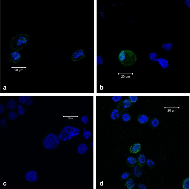
Figure 1.
Confocal microscopy of vitamin D receptor response (VDR) expression in human acute monocytic leukaemia cell line (THP1) cells. (a1 and a2) Confocal microscopy photos of non-stimulated THP1 cells labelled with VDR antibody and nuclear 4',6-diamidino-2-phenylindole (DAPI). VDR is present in some THP1 cells both in the nucleus and cytosol with different intensity. (b) Confocal microscopy photo of 25-hydroxyvitamin D3 (1·25-vitD)-stimulated THP1 cells labelled with isotype and nuclear DAPI. (c) Confocal microscopy photograph of 1·25-vitD-stimulated THP1 cells labelled with VDR antibody and nuclear DAPI. 1·25-vitD increases the VDR expression and induces a minor shift of VDR towards the nucleus compared to non-stimulated cells. Blue colour = nuclear DAPI; green colour = VDR.

