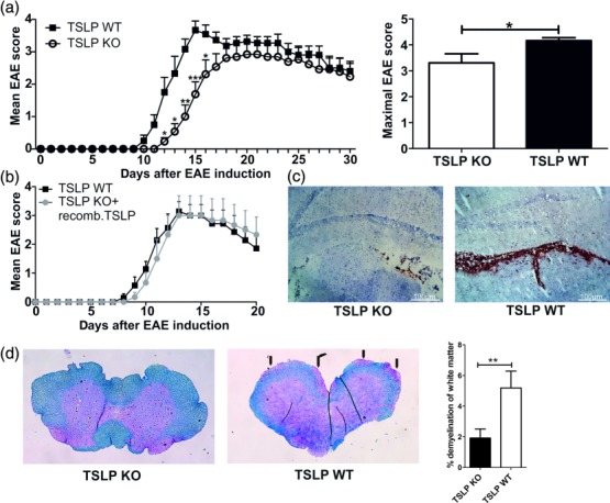
Figure 1.

Thymic stromal lymphopoietin (TSLP) knock-out (KO) mice show a delayed experimental autoimmune encephalomyelitis (EAE) development and a reduced EAE severity. (a) EAE was induced in male TSLP KO mice or wild-type (WT) control animals by immunization with myelin oligodendrocyte glycoprotein peptide 35–55 (MOG35–55) in complete Freund's adjuvant (CFA) and disease severity was monitored according to the classical EAE score [mean clinical EAE score ± standard error of the mean (s.e.m.); maximal clinical EAE score ± s.e.m.; TSLP KO n = 13, TSLP WT n = 12; data presented are the mean of three independent experiments; Wilcoxon–Mann–Whitney comparison test: *P < 0·05; **P < 0·01; ***P < 0·001]. (b) EAE was induced in TSLP KO mice and in TSLP WT mice. In addition, TSLP KO mice received rmTSLP [500 ng in 200 µl phosphate=buffered saline (PBS)] intraperitoneally (i.p.) on days 0–5; TSLP WT mice received 200 µl PBS i.p. on days 0–5; disease severity was monitored according to classical EAE score (mean clinical EAE score ± s.e.m.; TSLP KO n = 6, TSLP WT n = 7). (c) CD45 staining of brain harvested from TSLP KO and TSLP WT mice at day 12 after EAE induction. Results are representative of at least three independent experiments. (d) Luxol fast blue staining of spinal cord sections harvested from TSLP KO mice (left) and TSLP WT mice (middle) at day 12 after EAE induction; arrows mark demyelinated areas; bar graph (right) shows quantification of demyelinated areas (n = 8–10 mice; Wilcoxon–Mann–Whitney comparison test: **P < 0·01).
