
Figure 5.
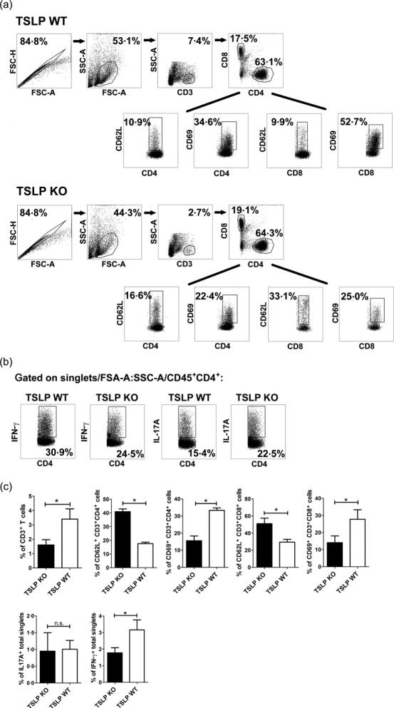
Fewer T helper type 1 (Th1) cells are present in brain of thymic stromal lymphopoietin (TSLP) knock-out (KO) mice at day 11 after experimental autoimmune encephalomyelitis (EAE) induction and infiltrating CD4+ and CD8+ T cells show an impaired activation. (a) CD45+ leucocytes isolated from pool of three brains were analysed for surface antigens CD4, CD8, CD62L and CD69 by flow cytometry. (b) Flow cytometric analyses of interferon (IFN)-γ and interleukin (IL)-17A production in singlets/forward-scatter (FSC)-A : side-scatter (SSC)-A/CD45+-gated CD4+ T cells. The figures (a) and (b) are representative of five independent experiments. (c) Statistical analyses by flow cytometric analyses of brain infiltrating total CD3+ T cells, CD69 and CD62L expression on CD4+ and CD8+ T cells, IL-17A and IFN-γ expression in brain singlets (bar graphs, n = 12–18; Wilcoxon–Mann–Whitney comparison test: *P < 0·05).