
Figure 6.
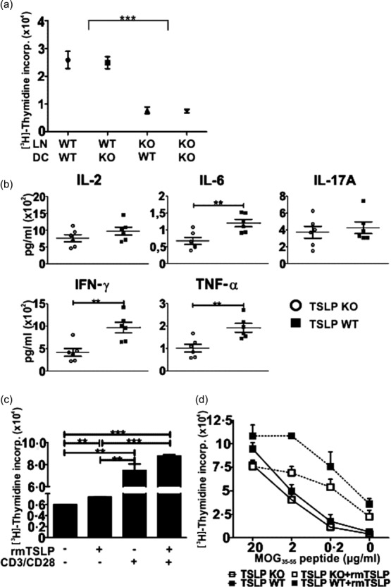
Thymic stromal lymphopoietin (TSLP) stimulates the proliferation of T cells. (a) 200 000 lymph node (LN) cells (Balb/c) were seeded together with 3000 tumour necrosis factor (TNF)-α-matured bone marrow dendritic cells (BMDCs) (C57/BL6) into a 96-well flat-bottomed plate for 72 h. Cell cultures were then pulsed with 1 μCi/well [3H]methylthymidine for 16 h and then the incorporation was assessed; TSLP KO n = 6, TSLP wild-type (WT) n = 6. (b) 200 000 KO or WT LN cells (Balb/c) were co-cultured with 3000 WT BMDCs (C57/BL6) for 72 h. Supernatants were then analysed regarding their cytokine concentrations by cytometric bead array (CBA); TSLP KO n = 6, TSLP WT n = 6. (c) LN cells from naive WT mice were seeded into a 96-well flat-bottomed plate (400 000/well) and cultured for 48 h in the presence or absence of CD3/CD28 beads and rmTSLP. Cell cultures were then pulsed with 1 μCi/well [3H]-methylthymidine for 16 h and incorporation was assessed; n = 3. (d) Splenocytes were harvested from TSLP KO and WT mice at day 5 after immunization with myelin oligodendrocyte glycoprotein peptide 35–55 (MOG35–55) peptide. CD3+ T cells were then isolated, seeded into a 96-well flat-bottomed plate (400 000/well) and restimulated for 72 h with different concentrations of MOG35–55 peptide in the presence or absence of rmTSLP. Cell cultures were then pulsed with 1 μCi/well [3H]-methylthymidine for 16 h and incorporation was measured; TSLP KO n = 6, TSLP WT n = 3. The bar and line charts represent the mean ± standard error of the mean. Results are representative of three independent experiments; two-tailed unpaired Student's t-test: *P < 0·05; **P < 0·01; ***P < 0·001.