
Figure 1.
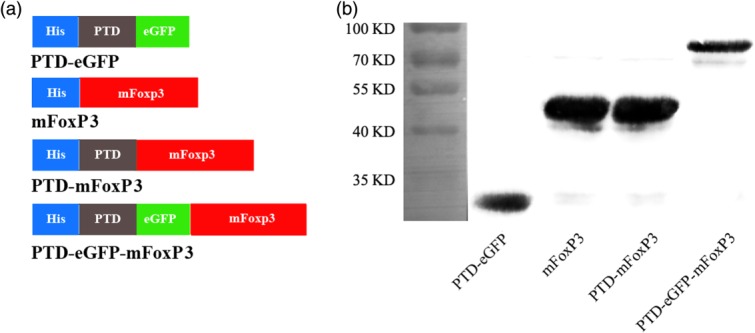
Preparation of the protein transduction domain (PTD) fusion proteins. (a) Schematic structures of the various recombinant proteins prepared and used in this study, including full-length mouse forkhead box protein 3 (mFoxP3), full-length mFoxP3 fused with the PTD sequence (PTD-mFoxP3) or with PTD plus enhanced green fluorescent protein (eGFP) (PTD-eGFP-mFoxP3) and a control PTD-eGFP. All the proteins were tagged a 6 × His sequence, represented by blue boxes. The grey box represents PTD peptide (YGRKKRRQRRR) derived from HIV-1 PTD protein. The green box represents an eGFP. (b) Western blotting analysis of purified recombinant proteins probed with mouse anti-6 × His Tag monoclonal antibody (mAb). Expected sizes of recombinant proteins were PTD-mFoxP3, 51 kDa; PTD-eGFP-mFoxP3, 80 kDa; mFoxP3, 50 kDa and PTD-eGFP, 33 kDa.