
Figure 1.
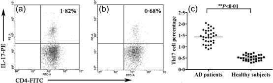
Representative images showing the T helper type 17 (Th17) cell percentage (CD4+IL17+/CD4+ T cells %). (a) A 16-year-old male atopic dermatitis (AD) patient; (b) a 17-year-old male healthy subject; (c) the Th17 cell percentage in peripheral CD4+ T cells in AD patients (n = 33) versus healthy subjects (n = 31).