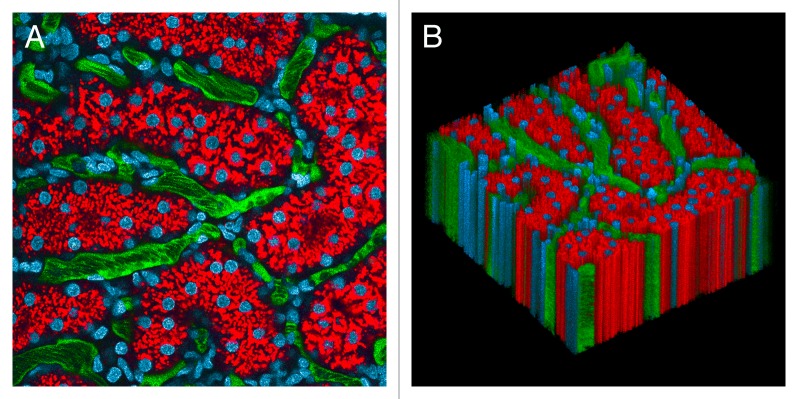
Figure 1. Intravital microscopy of the rat kidney mounted in a kidney cup. (A) First of 120 images collected from the kidney of a living rat, following intravenous injection of Hoechst 33342 (labels nuclei blue), 3000 MW TexasRed dextran (red, internalized into endosomes of proximal tubule cells) and 500 000 MW fluorescein dextran (green, in intertubular capillaries). (B) XYT volume rendering of the time series, with sequential images arrayed vertically in the volume. Image volume is 200 microns across. The time series and volume rendering are presented in Video S1.

An official website of the United States government
Here's how you know
Official websites use .gov
A
.gov website belongs to an official
government organization in the United States.
Secure .gov websites use HTTPS
A lock (
) or https:// means you've safely
connected to the .gov website. Share sensitive
information only on official, secure websites.
