
Figure 1.
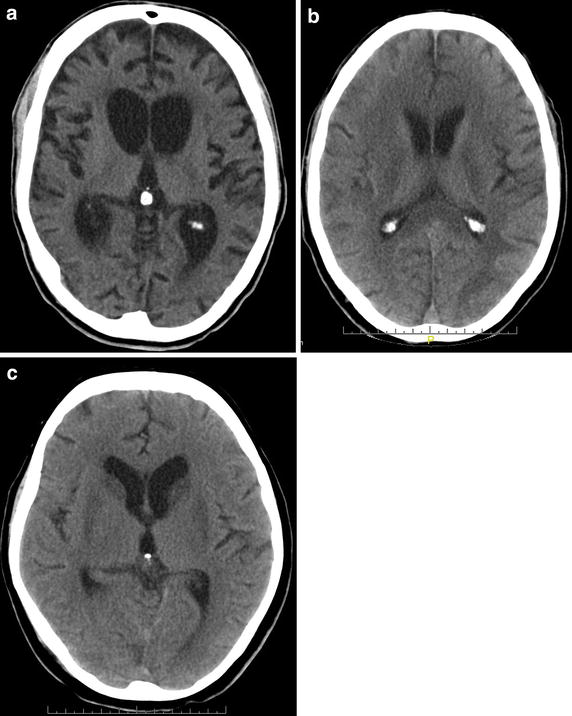
Typical CT scans of hypoxic brain damage patients. a Brain atrophy and hydrocephalus of a 44 y old male patient, 6 months after hypoxia. The patient was in a minimally conscious state (MCS). b Hypodense white matter changes of a 63 y old male patient, 2 weeks after hypoxic brain damage. The patient was in an unresponsive wakefulness syndrome (UWS). c Bilateral basal ganglia hypodensities of a 61 y old female patient, 6 weeks after hypoxic brain damage. The patient was in a MCS.