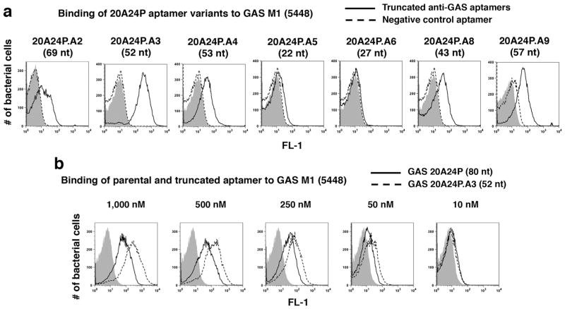
Fig. 2.
Truncation of GAS aptamer 20A24P from 80 to 52 nucleotides leads to enhanced binding to live GAS 5448 cells. a Binding of the indicated 5′-FAM labeled truncated versions of the GAS aptamer 20A24P (solid black lines) and 39-nt negative control aptamer RAND-39 (dashed lines) at 500 nM end concentration or aptamer vehicle (gray solid). For the data shown for 20A24P.A9, 3′-FAM labeled GAS aptamer (solid line) and 52-nt negative control aptamer RAND-52 (dashed line) were used. The length of the respective GAS aptamers is indicated in parentheses. b Binding comparison of parental 5′-FAM 80-nt GAS aptamer 20A24P (black solid lines) and 5′-FAM truncated 52-nt aptamer 20A24P.A3 (dashed lines) at the indicated aptamer end concentrations vs. aptamer vehicle (gray solid). For all samples, the bacteria were incubated with the aptamers or vehicle for 37 °C at 45 min, then washed and subjected to flow cytometry to measure the fluorescence intensities of 20,000 bacterial particles per sample. Each sample was run in duplicate with similar results

