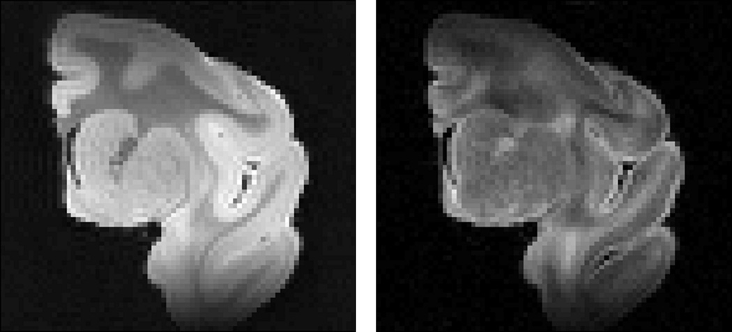
Figure 1.
Two-dimensional MRI coronal view of the macaque brain hemisphere used in this study. Left, nondiffusion-weighted “b0” image, b = 0 s/mm2. Right, diffusion-weighted image, b = 5000 s/mm2 in a single diffusion direction, scaled up for better display. There is notable shading due to coil inhomogeneity.

