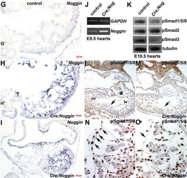
Figure 1.

Ectopic Noggin in the Nfatc1 endocardial lineage is embryonic lethal. (A–F): Resultant phenotype of NogEnd mutant embryos (B,D,F) compared to wildtype littermate controls (A,C,E) at E9.5, 10.5 and 12. Wholemount right sided views show that E9.5 and 10.5 NogEnd embryos are grossly unaffected but that E12 NogEnd mutants are runted and exhibit pooling of blood in both blood vessels (indicated via arrow in F) and heart itself when compared to wildtype control littermates; (G–I): Non-radioactive in situ hybridization analysis using an anti-Noggin DIG-labelled probe confirms absent endogenous mRNA expression in control E10 heart (G) but robust endocardially-restricted transgenic Noggin (blue signal) in NogEnd mutant heart within cells lining the atria, atrioventricular cushions and ventricle. G’ and H’ inserts represent low power images of whole heart sections; (J): Semi-quantitative RT-PCR measurement of Noggin levels in control and mutant E9.5 pooled hearts (n = 4 of each genotype) confirmed elevated Noggin expression levels (×6.4) in the mutant hearts (data shown from 34 PCR cycles). Note, GAPDH loading control (upper panel J) is similar in both samples (data shown from 20 PCR cycles); (K): Western analysis of TGFβ superfamily downstream effector Smad signaling in pooled E10.5 hearts (n = 4 of each genotype) revealed NogEnd mutant isolated hearts exhibit reduced pSmad1/5/8 levels (only ~35% of wildtype levels) when normalized to Tubulin levels, indicative of suppressed downstream BMP ligand signaling. Additionally, pSmad3 is similarly reduced in mutant hearts (only ~40% of wildtype levels), but pSmad2 levels remain unchanged. This suggests that downstream TGFβ ligand signaling is also partially suppressed; (L–O): Immunohistochemical analysis of pSmad1/5/8 in E10.5 control (L,N) and mutant (M,O) hearts demonstrates that expression is unaffected in cardiomyocytes but that pSmad1/5/8 expression (brown nuclear staining) is reduced in NogEnd mutant cushions compared to controls (* in (L)) and that pSmad1/5/8 expression is diminished within mutant endocardial cells (arrow in (O)) compared to wildtype controls (arrows in (N)). Scale bars: (G,H) = 20 μm; I = 50 μm. Abbreviations: lv, left ventricle; rv, right ventricle.

