Figure 3.
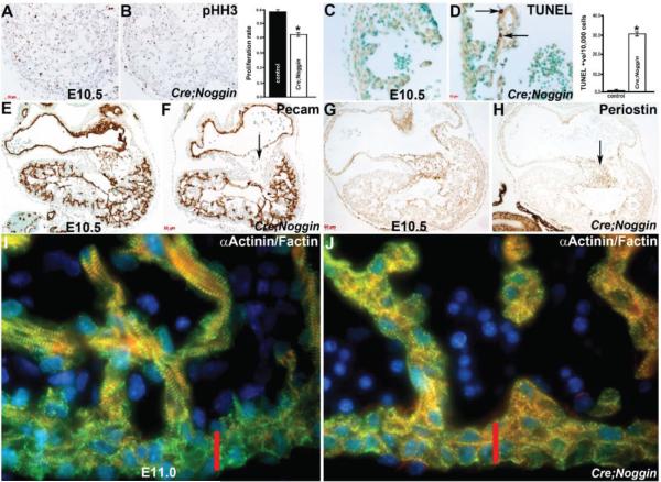
Immunohistochemical analysis of NogEnd mutant growth and development. (A,B): Phospho-histone H3 examination of cell proliferation revealed that cell proliferation is slightly reduced ~30% within E10.5 mutant hearts, as illustrated via reduced DAB-positive (brown) nuclear staining in mutant (B) when compared to control littermate (A). Quantitative analysis indicates that the proliferation rate in mutant hearts is significantly reduced compared with the control sample. Data were averaged from 3 independent embryos with error bars indicating SD. * p < 0.05 (Student t test); (C,D): TUNEL analysis revealed that E10.5 mutant (D) hearts exhibit apoptotic cells (arrows in (D)) within the trabeculae adjacent to the overlying lacZ-postive endocardium compared to zero apoptosis in control hearts (C); Quantitative analysis indicates that apoptosis is only observed in the mutant ventricle. Data were averaged from 3 independent embryos with error bars indicating SD. * p < 0.05 (Student t test); (E,F): Expression of the endothelial cell adhesion and signaling receptor PECAM-1 confirms that the endocardium is still present and intact within NogEnd heart atria and ventricles, and that the mutant endocardial cells overlying the hypoplastic cushions (arrow in (F)) is unaffected. Note that the weaker PECAM-1 immunoreactivity in the control cushion is non-specific background staining, as endocardial cells undergoing EMT exhibit minimal to no pECAM-1 staining; (G,H): Reduced expression of the TGFβ -responsive endocardial cushion marker Periostin indicates that mutant endocardially-derived cushions are anomalous (arrow in (H)) compared to control littermates (G); (I,J): Confocal immunofluorescent imaging of F-actin (red), Actinin (green) with DAPI nuclear staining (blue), reveals that while these cytoskeletal proteins are present within both control (I) and mutant (J) hearts, there are far fewer mature contractile fibrils within the E11 NogEnd mutant cardiomyocytes when compared to littermate controls. Moreover, the normal stratification of mature contractile apparatus within primarily the trabeculae rather than compact zone cardiomyocytes (indicated via red bar) observed in control heart (I) is not present in NogEnd mutant hearts (J); Scale bars: (A,B) = 50 μm; (C,D) = 10 μm; (E–H) = 20 μm.

