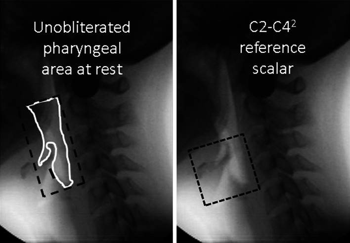
Fig. 1.
Lateral videofluoroscopic images taken after the hyoid has returned to rest. The left-hand image shows a tracing of the unobliterated pharyngeal area in white, with the boundaries used for pharyngeal area tracing shown in the dashed black line. The right-hand image illustrates the anatomical reference scalar used for normalizing measures of pharyngeal area. Normalized area measures can be interpreted as reflecting a percentage of this scalar reference area

