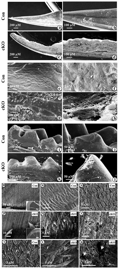
Fig. 4.
SEM of the mandibular incisors and molars from Bmp2 null mice. a. Enamel layer of the incisor of a 3 month-old control mouse is smooth. b, e and f. Higher magnification of a. c. Enamel layer of the incisor at a 3 month-old Bmp2 cKO mouse is rough. The buccal surface is jagged and tip of the incisor is abraded on the enamel layer. d, g and h. Higher magnification of c. i. Molar of a 3 month-old control mouse. k. Molar cusps in the null mouse are rugged and abraded. j and l. Higher magnification of i and k. m, n, o and s. Enamel in the control mice shows a typical prismatic pattern of rod (r) and inter-rod (ir) structures. Rods and inter-rods are well aligned. q, r, t and u. Enamel in the out layer of the null mice is abraded and has irregular architectures of rods and inter-rods. The rods and inter-rods have disorganized prismatic patterns. The broken surface of the enamel between the rods and inter-rods forms numerous “holes” (arrows) ranging from about 1 to 10 μM in sizes. The transverse section from the control mice in the panel m is shown in inset. The transverse fracture from Bmp2 cKO mice in the panel p is shown in inset. Bar, 100 μM.

