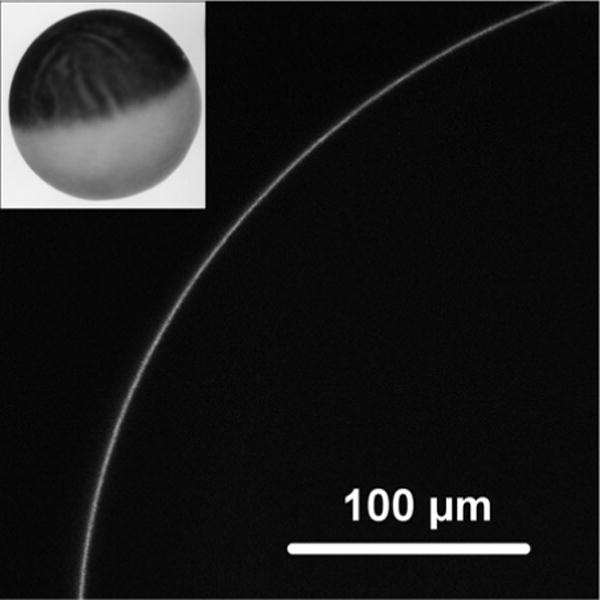
Fig. 5.

A laser-scanning confocal micrograph of the fluorescent signal from the periphery of an oocyte RNA 7 days after injection with MscS-GFP cRNA. An oocyte in ND96 buffer was placed onto a glass slide with concaved bottom and covered with a coverslip. Under bright field the lens was focused on the edge of the oocyte, then the confocal scan with GFP filters (488/510 nm) setup was performed. Inset: oocyte image in bright field
