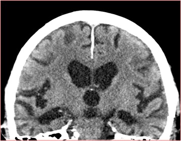
Fig. 1.

Medial temporal lobe atrophy. Example of abnormal medial temporal lobe atrophy in a CT scan in a study patient representing a score of 3 on the left side and 4 on the right side. This was not mentioned in the original report. This patient had an MMSE score of 22 points with 0 points on the memory item. This patient had noprevious mentioning of cognitive impairment in medical records
