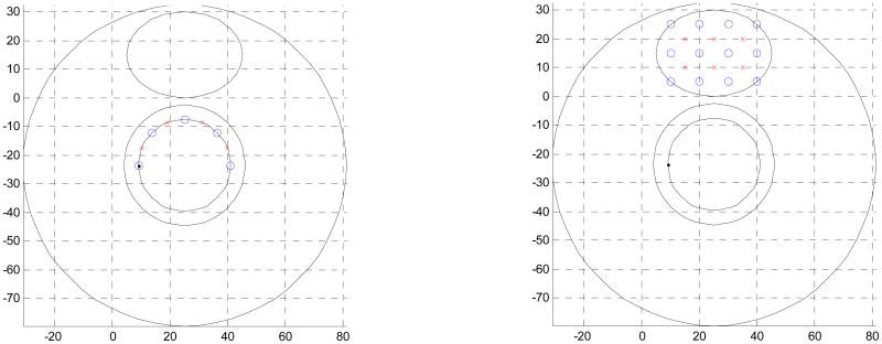
Figure. 1.
Schematic of endoscopic and interstitial source-detector geometry cross-sections used in prostate diffuse optical tomography applications. Endoscopic geometry (a) has been reported being used non-invasively in the canine experiments, while the interstitial geometry has been used for light dosimetry during PDT treatment. Blue circles are the detector positions, the red crosses are the source positions.

