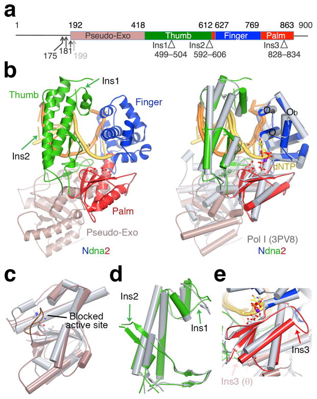
Figure 1.
Structure of Pol ν. (a) The primary structure. The N-termini of four Pol ν variants are indicated by arrowheads. Structural domains and three insertions are marked with boundary residues. (b) Ndna2 structure (left) and its superposition with bacillus Pol I 33 (grey, right). DNA is colored yellow (primer) and orange (template). The catalytic residues and incoming nucleotide in Pol I are shown as ball–and–sticks. (c) A zoom-in view of the superimposed Exo domain. Pol ν Exo site is mutated (equivalent residues in bacillus Pol I are shown in sticks) and blocked by a long loop. (d) The Thumb domain. (e) The Palm domain. Ins1–3 are marked by arrowheads (d,e).

