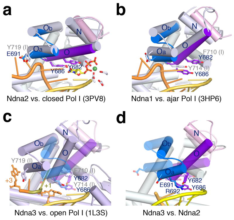
Figure 2.
The unusual finger-open structure of Pol ν75. (a) Superposition of Ndna2 (protein only with helices N, O, Oa and Ob in different colors) and the finger-closed Pol I (3PV833, grey protein, yellow primer and orange template). Key residues are diagramed. (b) Superposition of Pol ν of Ndna1 and the finger-ajar Pol I with DNA (3HP635). (c) Superposition of Ndna3 (semi-transparent Pol ν) with the finger-open Pol I with DNA (1L3S34, light blue). (c) Superposition of Ndna3 (protein and DNA) and Ndna2 (semi-transparent). Rotations of helices in Ndna3 relative to Pol I and Ndna2 are indicated by red and yellow arrows.

