
Fig. 5.
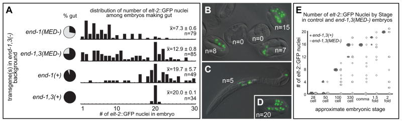
Number of gut nuclei becomes highly variable when gut specification is partially compromised. (A) Histograms showing relative number of embryos with amount of elt-2::GFP nuclei as indicated along the bottommost X-axis; only embryos containing at least one elt-2::GFP nucleus are included. The relative bar heights are scaled to the highest numeric class for each case. To the left of the histograms, a pie chart shows the proportion embryos containing gut without regard to number of gut nuclei. The reporter itself does not cause the changes in gut nuclei, as similar strains lacking the reporter still exhibit patches of gut granules (e.g. many embryos Fig. 3E), and we were able to see similar effects on number of nuclei expressing an integrated pept-1::mCherry reporter (not shown). (B–D) DIC micrographs digitally overlaid with GFP fluorescence. (B) Example of late-stage end-1,3(MED-) embryos with no gut and variable amounts of gut nuclei shown by elt-2::GFP expression. (C) A terminal L1 larva showing four nuclei in the anterior and one in the posterior. Note presence of gut granules (white speckles) around these nuclei. (D) Appearance of elt-2::GFP in a late-stage control embryo. (E) Number of elt-2::GFP-expressing nuclei at various stages during embryonic development, comparing the control strain (solid circles) with the end-1,3(MED-) strain (open circles).