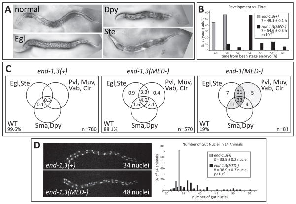
Fig. 6.
Post-embryonic defects are apparent in specification-compromised strains. (A) Phase-contrast images of adults on plates. (B) Bar chart showing that relative to controls, surviving adults of the end-1,3(MED-) strain experience an average developmental delay of 5.5 h during larval development. 134 control and 90 end-1,3(MED-) animals were scored. (C) Venn diagrams showing appearance of phenotypes as a percentage of total number of adults scored. Within the circles, regions are shaded gray in proportion to the percentage. Egl = egg laying defective; Ste = sterile; Pvl = protruding vulva; Muv = multivulva; Vab = variable abnormal; Clr = clear, translucent appearance; Sma = small body size; Dpy = dumpy body shape; WT = wild-type (normal) appearance. (D) Number of gut nuclei varies in late L4 stage animals. The images show sample fluorescence of a pept-1::mCherry::H2B reporter in each of the two strains. The bar chart shows that surviving end-1,3(MED-) L4-stage animals have a variable number of gut nuclei and a higher mean overall. 24 control and 50 end-1,3(MED-) animals were scored. Anterior is to the left in panels (A) and (D), which are also at the same scale. The normal adults are approximately 1mm long.

