
Figure 2. Csf1r-GFP+ PM macrophages make contact with Flk1-myr::mCherry+ endothelial cells and engulf endothelial cell plasma membrane.
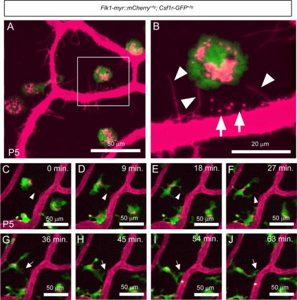
Static imaging at P5, showed Cherry+ plasma membrane particles localized to GFP+ macrophages (A). Higher magnification of the boxed area in panel A shows the PM endothelium as sending out cytoplasmic extensions which project toward the macrophages (arrowheads in B) as well as Cherry+ puncta localized outside of the vasculature (arrows in B). Live imaging of the PM at P5 confirmed that macrophage filopodia make intermittent contact with the Cherry+ cytoplasmic extensions (arrowheads in C to F). Macrophage filopodia also make contact with the walls of the PM vasculature and appear to “grab” plasma membrane particles from the vessel surface (arrows in G to J). N>5.