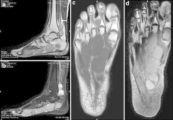
Figure 2.

Magnetic resonance imaging of the right foot and ankle. a Sagittal T2-weighted image showing multiple hyperintense lesions along the course of the posterior tibial (white arrow) and medial plantar nerves. b Sagittal T2-weighted image with fat suppression demonstrating multiple nodular lesions with heterogeneous high signal intensity. c Coronal T1-weighted image showing the lesions with iso-signal intensity relative to adjacent muscle. d Coronal contrast-enhanced T1-weighted image with fat suppression revealing mild to moderate enhancement of the lesions.
