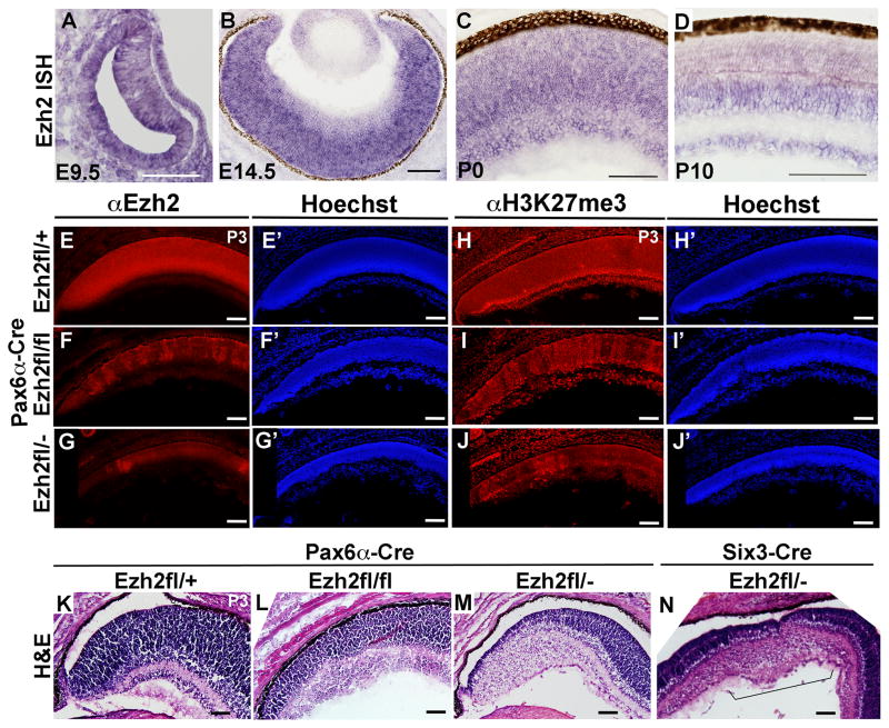
Figure 1. Ezh2 expression during retinal development and after Ezh2 conditional deletion.
(A–D) In situ hybridization (ISH) analysis. (E–G) EZH2 immunostaining shows increasing loss of protein in the peripheral retina with conditional deletion using Pax6-αCre in Ezh2fl/fl and Ezh2fl/− mice relative to control Ezh2fl/+ mice at P3. (E′–G′) Hoechst staining of the same sections. (H–J) Similarly H3K27me3 immunostaining shows increasing loss of this histone mark. (H′–J′) Hoechst staining of the same sections. (K–M) Hematoxylin and eosin staining shows increasing disruption to retinal lamination with conditional deletion using Pax6-αCre in Ezh2fl/fl and Ezh2fl/− mice relative to control Ezh2fl/+ mice at P3. (N) Similar disruption of retinal lamination in the central retina is observed after conditional deletion of Ezh2 using Six3-Cre in Ezh2fl/− mice. Scale bar = 100μm

