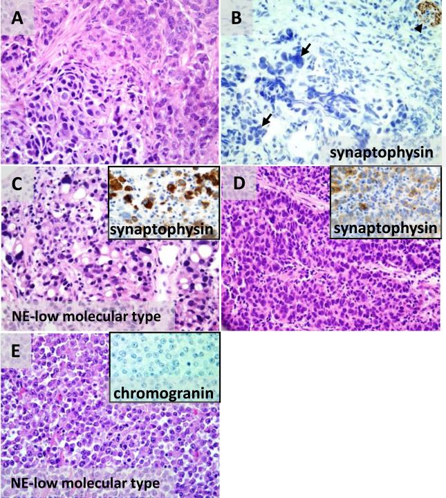
Figure 7. Agreement between neuroendocrine differentiation by morphology and immunohistochemistry is imperfect.
(A-B) A tumor with two distinct neuroendocrine cytologic patterns on H&E, salt-and-pepper chromatin (A, upper right) and hyperchromasia and nuclear molding (A, lower left) is negative for synaptophysin (B, arrows at tumor cells with crush artifact) with positive internal control synaptophysin staining in the stromal nerve (B, arrowhead). The tumor cells are also negative for chromogranin protein (not shown). (C-D) An NE-high molecular subtype carcinoma has spatially separate zones of pleomorphic signet ring cells (C) and trabecular/solid nests of non-signet ring cells with high grade neuroendocrine morphology (D). In both zones the malignant cells express synaptophysin (insets in G and H). (E) A carcinoma of NE- low molecular type and solid histology has well-defined cellular borders, prominent nucleoli, absent nuclear molding, and sheet-like growth, and is negative for chromogranin (inset) and synaptophysin (not shown).

